

Lanthanide-Based Imaging Probes
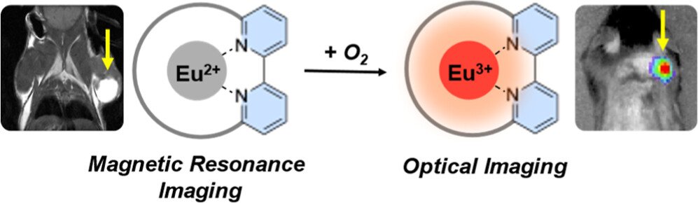
The prognosis and survival of patients with aggressive cancers depends on the presence of positive tumor margins (defined as the presence of tumor cells in the surrounding area) post-surgical resection. Combining radioactive and luminescent reporters in a targeted molecular probe has the potential to provide pre-operative nuclear imaging, real-time luminescence-guided surgery, followed by ex vivo imaging with one single probe. We are interested in employing luminescent lanthanides for in vivo optical imaging, a thus far insurmountable challenge due to the lanthanides’ need for short wave excitation. We have recently bypassed the need for short-wave, external excitation sources by carrying out in situ excitation of lanthanide luminescence with Cherenkov radiation emissive radioisotopes. Following extensive in vitro validation, we currently work on demonstrating that lanthanides are suitable for the in vivo optical imaging of cancer.
Representative publications:
Rodgers, C. B., Deal, M. P., Garman, L. C., Guzei, I. A., Allen, M. J., Boros, E. “Single Molecule Eu 2+/3+ Complex Platform for Optical and Magnetic Resonance Imaging in Vivo”. J. Am. Chem. Soc. 2026, 148, 23, 24520–24530.
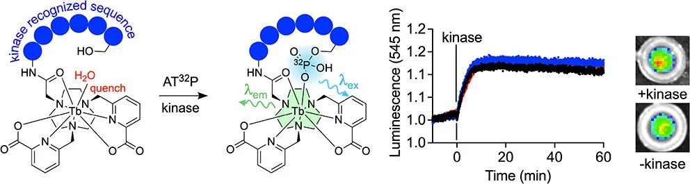
Sands G., Lao Y., Joaqui-Joaqui M.A., Huang X., Boros E., “Accessing Self-Illuminated, Luminescent Lanthanide Probes by Enzymatic Radiophosphorylation” Inorg. Chem. 2025, 64, 51, 25270–25280.
R. Lengacher, K. E. Martin, D. Śmiłowicz, H. Esseln, P. Lotlikar, A. Grichine, O. Maury, E. Boros. “Targeted, Molecular Europium(III) Probes Enable Luminescence-Guided Surgery and 1 Photon Post-Surgical Luminescence Microscopy of Solid Tumors” J. Am. Chem. Soc. 2023, 145, 44, 24358–24366
A. G. Cosby, G. Quevedo, and E. Boros. A High-Throughput Method To Measure Relative Quantum Yield of Lanthanide Complexes for Bioimaging. Inorg. Chem. 2019, 58, 10611-10615